Our center operates two 3 Tesla scanners for versatile research applications, as well as a 7 Tesla ultra-high-field system that enables ultra-high-resolution imaging of fine-scale anatomy, microstructure, and functional organization. MRI supports a broad range of methods, including structural imaging, functional MRI (fMRI), diffusion tensor imaging (DTI), perfusion imaging and quantitative techniques (qMRI) for studying brain connectivity and tissue properties. These approaches are used to investigate neuroanatomy, cognition, neurological and psychiatric conditions, and biomarkers of brain health.
Magnetic Resonance Imaging (MRI)
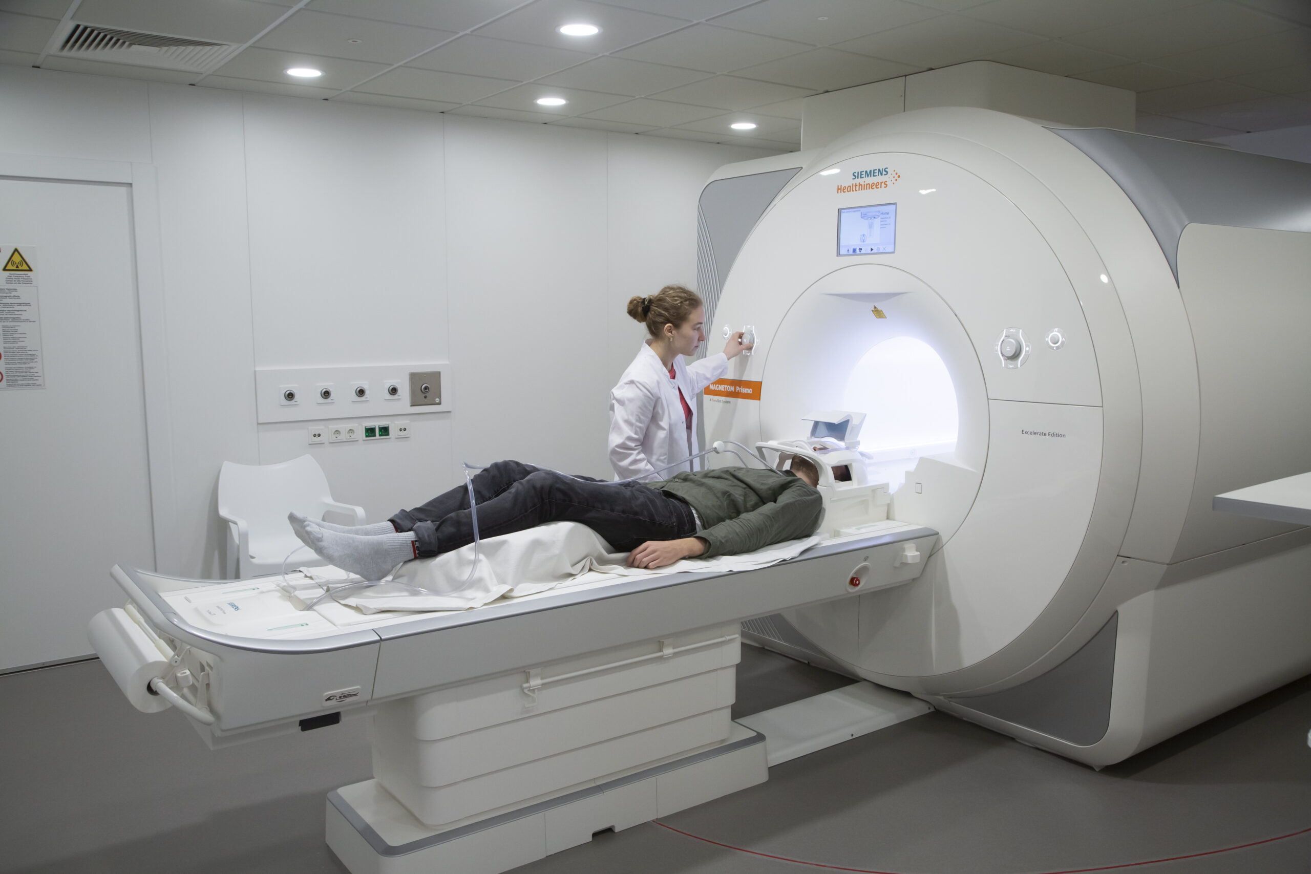
MRI allows us to visualize brain structure and function in exquisite detail using strong magnetic fields and electromagnetic waves.
During a scan
During an MRI scan, participants lie on a padded table that slides into the scanner bore and are asked to remain still while images are acquired. Certain types of equipment are not allowed inside the scanner room, such as metallic objects, credit cards, active body implants or electrical devices. The procedure is non-invasive and does not involve ionizing radiation. The scanner produces acoustic noise at different frequencies, so hearing protection is provided. Depending on the protocol, participants may be asked to rest or perform simple tasks while visual or auditory stimuli are presented. Trained staff remain in continuous contact throughout the session. We screen every participant before entering the MR room for MR contraindications like implanted pacemakers to ensure subject safety. Some people may experience short-lasting vertigo when being moved into or out of the scanner bore, especially at 7T, or peripheral nerve stimulation during scanning, or may find the scanner bore narrow. Otherwise, the procedure is completely harmless.
Electroencephalography (EEG)
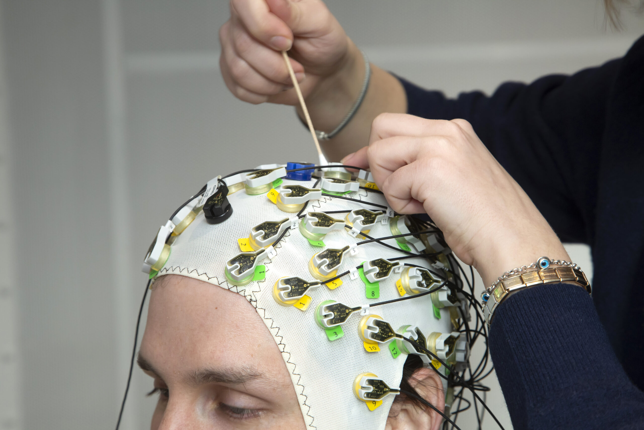
Electroencephalography (EEG) records electrical brain activity from electrodes placed on the scalp, offering a flexible and widely applicable method for studying neural function.
EEG provides excellent temporal resolution and is well suited for investigating sensory processing, cognition, sleep, and brain network dynamics. It can be used independently or in combination with MRI and MEG to enable multimodal analyses of brain activity. EEG is also valuable for longitudinal studies and experimental paradigms requiring portable or extended recordings.
During an EEG session
During an EEG session, participants wear a lightweight cap fitted with small sensors that detect electrical signals from the brain. The setup is painless and non-invasive. Tasks may include viewing images, listening to sounds, or resting quietly. Our staff guide participants through each step of the process and ensure comfort throughout the recording.
Magnetoencephalography (MEG)
When thousands of nerve cells fire synchronously, they generate tiny, precisely timed magnetic fields – about a billion times weaker than a refrigerator magnet.
Inside the MEG helmet, superconducting quantum interference devices (SQUIDs) cooled to –269°C detect these fields millisecond by millisecond. Using advanced algorithms, their cortical origins can be localised with an accuracy of a few millimetres. MEG is the most precise non-invasive method for directly recording neural activity.
This unprecedented temporal resolution makes MEG the ideal tool for researching cognitive and neural dynamics – the moment-by-moment choreography of perception, emotion, memory, language and action. Modern analysis techniques – including machine learning, multivariate pattern analysis and network modelling – now make it possible to visualise the millisecond-precise development of distributed neural activity patterns across cortical networks.
Whether it’s deciphering the rapid processing chain from sound to meaning, tracking the neural signatures of action planning and execution, or investigating anticipatory activity patterns prior to an expected event, researching cognitive and neural dynamics requires temporal precision in the millisecond range. And that’s exactly what MEG was developed for.
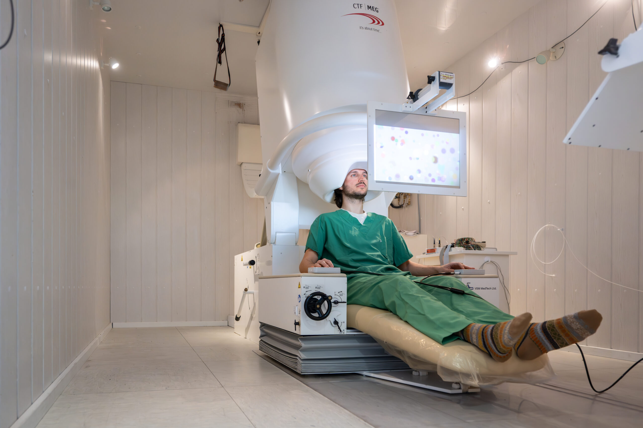
Our laboratory is equipped with a CTF whole-head MEG system with 275 axial gradiometer channels. It is located in a magnetically shielded room and is combined with high-resolution binocular eye tracking and visual, auditory and tactile stimulation equipment. The facility is open to both member institutions of the Cooperative Brain Imaging Centre and external users. The MEG team ensures high-quality data collection and provides training and support to scientists at all levels of experience.
During MEG
Participants undergoing MEG sit comfortably beneath a helmet-shaped sensor array while brain activity is recorded. The measurement is completely non-invasive and silent, and typically involves resting quietly or performing simple computer-based tasks. Head position is monitored to ensure data quality, and sessions are supervised by experienced operators. No electric or magnetic equipment is allowed inside the shielded room. Participants are in contact with the staff that is sitting directly in front of the room. The environment is designed to be calm and supportive, allowing participants to relax while high-quality neural data are acquired.
Transcranial Magnetic Stimulation (TMS)
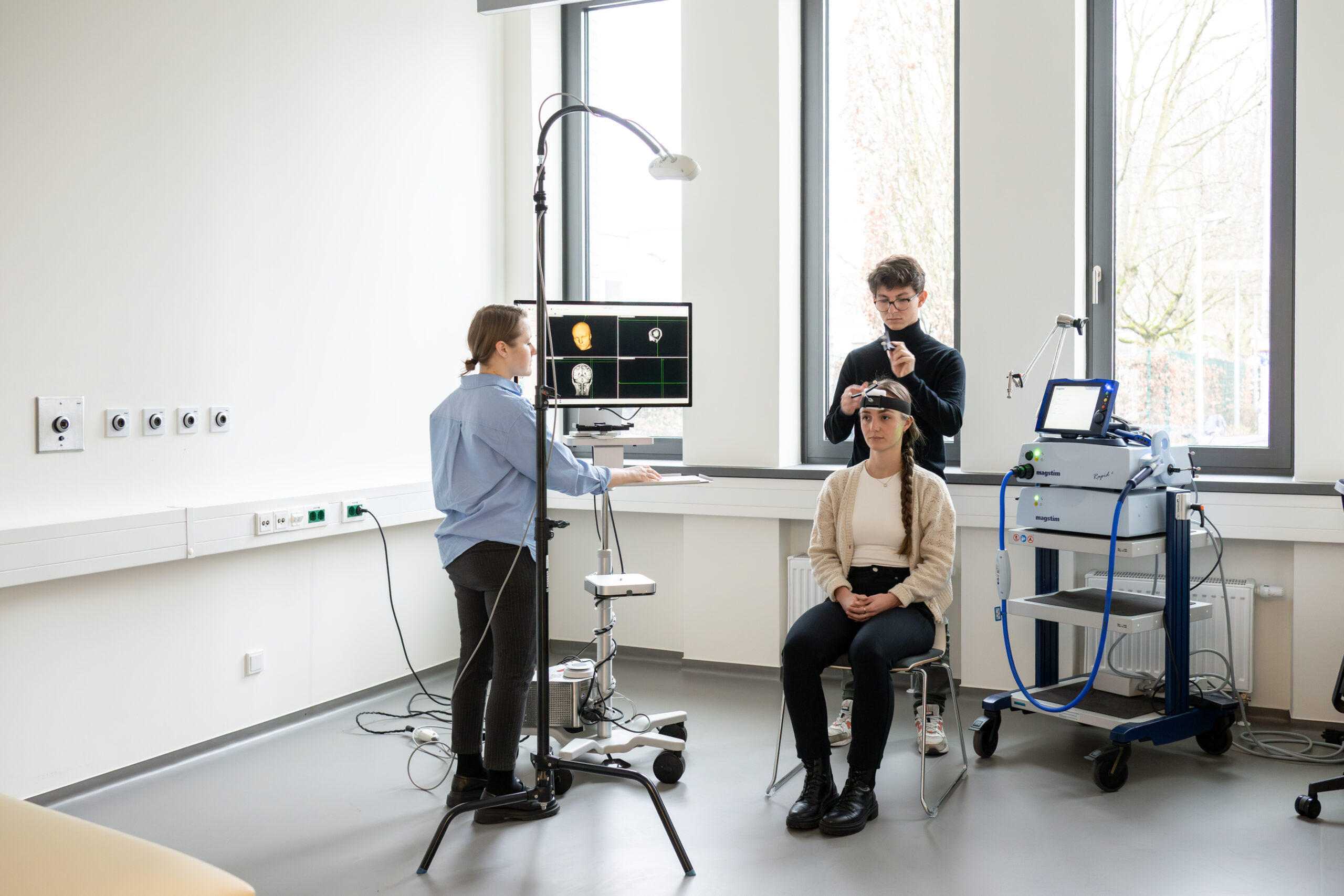
TMS is a non-invasive neuromodulation technique that uses brief magnetic pulses to stimulate targeted regions of the brain.
TMS allows us to probe causal relationships between brain activity and behavior, complementing observational methods such as MRI, MEG, and EEG. It is widely used in cognitive neuroscience to study functional brain networks. At our center, TMS can be combined with neuroimaging and electrophysiology to assess both immediate and longer-term effects of stimulation.
During TMS
During TMS, a coil is placed gently against the scalp, delivering short magnetic pulses to specific brain areas. Participants may feel a light tapping sensation, and occasional muscle twitches are normal. Sessions are conducted under established safety guidelines and supervised by trained personnel. Stimulation parameters are individualized, and participant comfort is carefully monitored at all times.